| Technical Reports | Work Products | Research Abstracts | Historical Collections |
![]()
|
Research
Abstracts - 2006
|
|
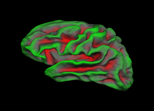
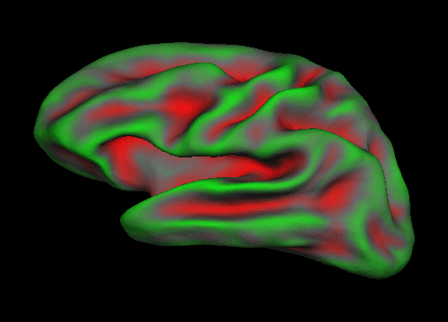
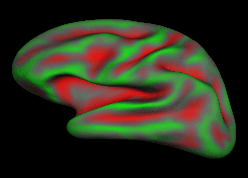
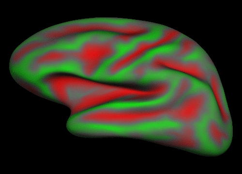
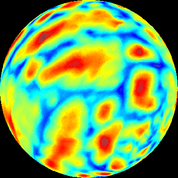
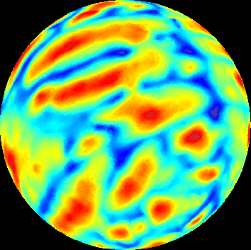
Modeling Cortical SurfacesBoon Thye Thomas Yeo & Polina GollandGoalWe are interested in developing techniques for analyzing the shape and folding pattern of cortical brain surfaces. In particular, we are comparing and analyzing the cortical patterns of neonates and young adults. Neurobiologists believe that most of our higher cognitive abilities originate from the cerebral cortex, and that neurological growth or diseases significantly alter the structure of the cortex. Our long term goal is therefore to find useful representations or features of cortical surfaces that allows us to track changes and development of the brain (and indirectly cognitive capabilities). As an ultimate goal, we would like to generalize such techniques to general object shapes. Proposed ApproachBecause cortical surfaces are highly convoluted structures, an approach commonly used in medical vision is to inflate and project the surfaces onto the sphere (see figure below) [1,2,3,4].
The resultant spherical images of the cortical surfaces are more easily manipulated albeit being incomplete representations of the cortex. The figure below shows examples of spherical representations of cortical surfaces. The colors correspond to "normalized depth", which is a measure of the depth and height of the sulci and gyri of the brain. The red color corresponds to sulci and the blue color corresponds to gyri.
In most literature, 2D non-linear registrations of the surfaces are then performed. Pixel correspondence is then assumed and an entire battery of tests (such as t-tests, wavelet analysis and jacobians of the deformations) is then applied. However, due to the variability of the brain surfaces, it is not clear whether the assumption of pixel correspondence after registration is valid. For example, it is wrong to claim pixel correspondence between a brain with one central sulcus versus one with two central sulci because there does not exist a diffeomorphism between the surfaces. Furthermore, tests of statistical significance yield results in the form of "significant pixels", which are unsatisfying since they are unintuitively related to the neuroanatomy. In contrast, we propose to avoid establishing correspondences at the pixel level and instead model the cortical folding pattern through a graphical model whose nodes correspond to whole sulci. The correspondences are then established at the level of hidden variables and hyperparameters. Differences between populations (e.g. young vs old, sick vs normal) in the values of the model parameters will provide a more intuitive interpretation of anatomical differences than pixel-wise methods. Collaborators (in alphabetical order)Bruce Fischl (MGH), Ellen Grant (MGH), Rudolph Pienaar (MGH) AcknowledgementThis work is funded in part by the NIH grants R01-NS051826 and 1U54 EB005149. Boon Thye Thomas Yeo is funded by Agency for Science, Technology and Research, Singapore. References:[1] Dale, A.M., Fischl, B. and Sereno M.I., Cortical surface-based analysis. I. Segmentation and surface reconstruction, Neuroimage, (9):174-194, 1999. [2] Fischl, B., Sereno M.I. and Dale, A.M., Cortical surface-based analysis. II: Inflation, flattening, and a surface-based coordinate system, Neuroimage, (9):195-207, 1999. [3] Tosun, D. and Prince, J.L., Mapping techniques for aligning sulci across multiple brains, Medical Image Analysis, (8):295-309, 2004. [4] Tosun, D. and Prince, J.L., Cortical Surface Alignment Using Geometry Driven Multispectral Optical Flow, Information Processing in Medical Imaging (IPMI), 2005 |
|||||||||||||||||
|
||||||||||||||||||