| Research Abstracts Home | CSAIL Digital Archive | Research Activities | CSAIL Home |
![]()
|
Research
Abstracts - 2007
|
|
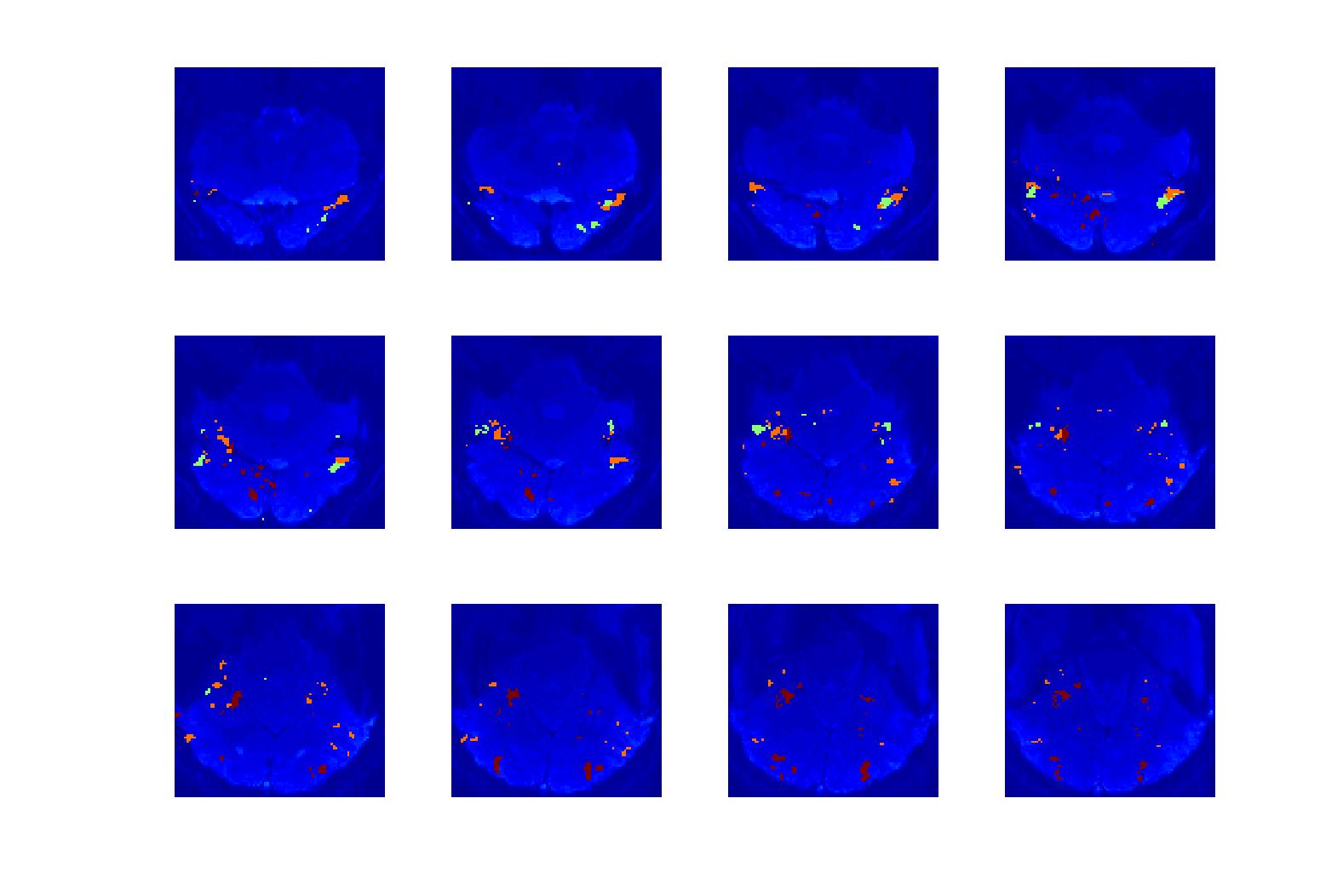
Algorithms for fMRI Study of High Level VisionDanial Lashkari & Polina Golland
BackgroundToday, functional Magnetic Resonance Imaging (fMRI) data is being extensively used for the investigation of a wide variety of different cognitive and perceptual brain processes. In particular, there exists comprehensive literature on the study of high level vision based on the fMRI data. The central idea is using fMRI data to understand if there are regions in the brain more responsive to certain high level categories of visual images, and the possible networks of such regions that enables the brain to classify its visual perception of the environment [1]-[3]. Currently, the conventional method for fMRI data analysis is based on statistical tests comparing response of each voxel in the brain to different visual categories to detect if it shows considerably higher activation to one category. In particular, the activations of a particular voxel when presented with different categories (e.g., faces and objects), are tested against a null hypothesis of both activations being from the same Gaussian distribution. If the null hypothesis is rejected with high significance, we consider that voxel to be included in a region corresponding to the preferred category. For example, the well-known FFA (Fusiform Face Area) is the set of voxels which show high activation to faces when compared to objects [4]. Formulation of the ProblemAlthough the described method provides us with relatively robust results, it can only be used to detect regions that show high response to specific parts of the stimulus. In this project, we are considering applications of more sophisticated learning methods to the fMRI images from such experiments with the goal of gaining more information about the underlying high-level visual perception network in the visual cortex. The ultimate goal of such a learning approach to the fMRI data of high level vision is to identify the visual classes and their (possible) corresponding brain regions, and understanding the structure of the connections between these classes and regions. A problem stated in this way can be answered through an algorithm which considers different possible mutual relationships between the temporal-spatial dimensions of data, as well as different possible structures in which they interact with each other. As the first stage, we are applying unsupervised clustering algorithms to detect regions especially interested to specific types of visual stimuli without introducing the regular statistical testing ideas. An algorithm for the biclustering of brain voxels and visual stimuli will enable us to further investigate the potential for learning from fMRI data in this framework. CollaboratorsProfessor Nancy Kanwisher Group, Department of Brain and Cognitive Sciences, MIT Research SupportThe National Alliance for Medical Image Analysis (NIH NIBIB NAMIC U54-EB005149)
References[1] N. Kanwisher, et al, Functional imaging of human visual recognition, Cognitive Brain Research, 5, pp.55--67, 1996. [2] N. Kanwisher, Domain specificity in face perception, In Nature Neuroscience, 3, pp. 759--763, 2000. [3] J. V. Haxby, et al, Distributed and overlapping representations of faces and objects in ventral temporal cortex, Science, 293, pp. 2425--2430, 2001. [4] N. Kanwisher, et al, The Fusiform Face Area: a module in human extrastriate cortex specialized for face perception, The Journal of Neuroscience , 17, pp. 4302--4311, 1997. |
|||
|