
| Research Abstracts Home | CSAIL Digital Archive | Research Activities | CSAIL Home |
![]()
|
Research
Abstracts - 2007
|
|
Stochastic Tractography and Applications in SchizophreniaTri M. Ngo, Marek Kubicki, Carl-Fredrik Westin & Polina GollandAim/Goal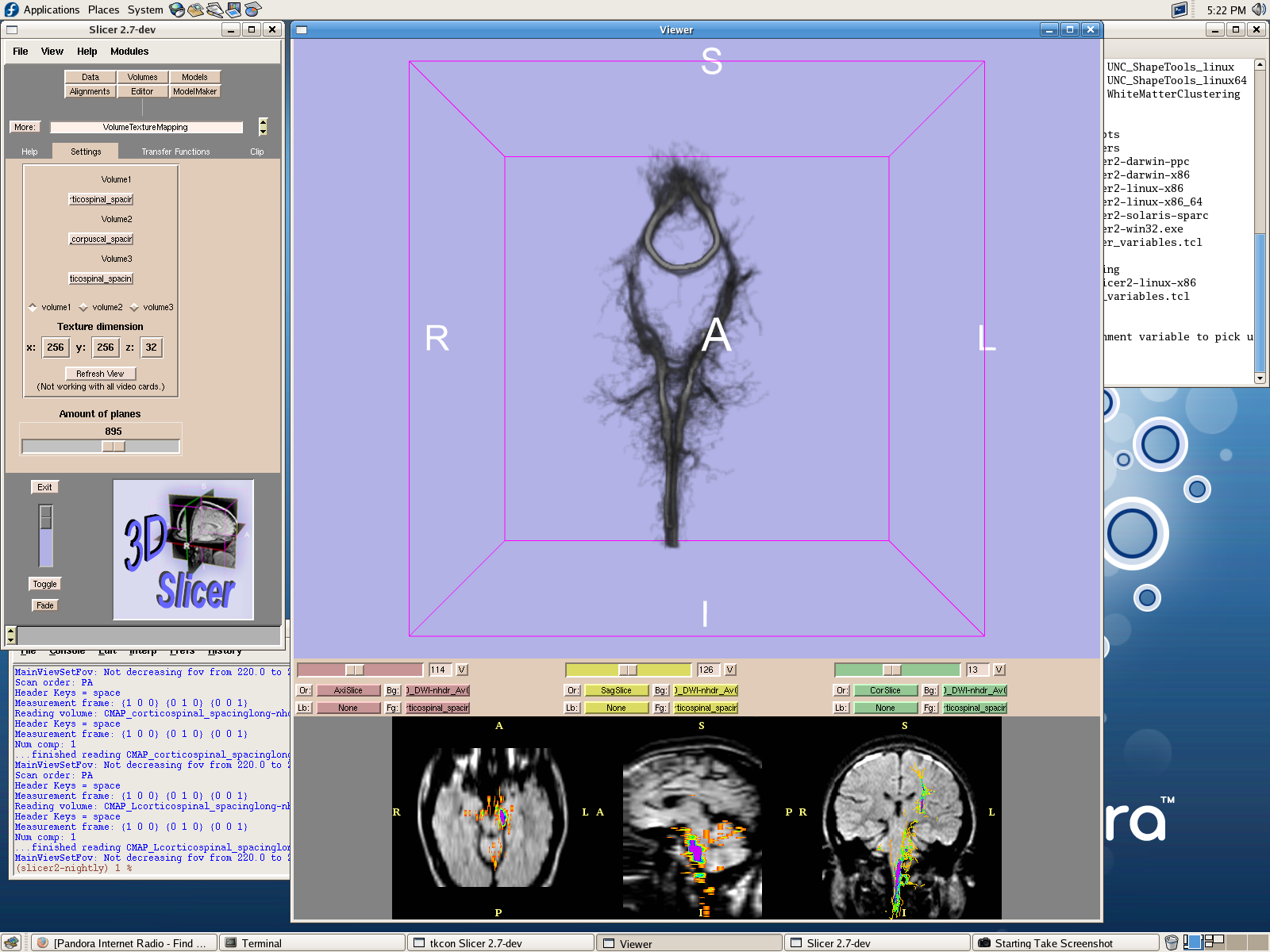
A connectivity map generated using the stochastic tractography algorithm. A single seed point is located in each of the left and right corticospinal tracts and in the corpus callosum. Stochastic Tractography is a Bayesian approach to estimating nerve fiber tracts from DWMRI (Diffusion Weighted Magnetic Imaging) images. The Bayesian framework provides a measure of confidence regarding the estimated tracts. This measure of confidence allows the algorithm to generate tracts which pass through regions with uncertain fiber directions, revealing more details about structural connectivity than non-Bayesian tractography algorithms. We will further develop the theory and application of stochastic tractography by applying it in a clinical study of nerve fiber tract abnormalities in schizophrenia. BackgroundMagnetic Resonance Imaging (MRI) is a valuable imaging modality for studying the brain in-vivo. We can use use MRI to differentiate between tissue types, which is valuable for anatomical studies. However, anatomical MRI provides a homogeneous image of white matter making it difficult to characterize white matter fiber tracts which pass through this region. Diffusion Weighted Magnetic Resonance Imaging (DWMRI) provides information about the diffusion of water molecules in the brain. DWMRI images can be used to construct a DTI data set which provides a complete description of water diffusion. Researchers have hypothesized that white matter abnormalities may underlie some neurological conditions. For instance, the neurological disease schizophrenia by is characterized by its behavioral symptoms, which include auditory hallucinations, disordered thinking and delusion[6]. Studies have suggested that these behavioral symptoms have some connection with the neuroanatomical abnormalities observed in schizophrenia patients. Using DTI, Researchers can noninvasively investigate the relationship between brain white matter abnormalities and schizophrenia by using DTI. We can visualize DTI data sets using a number of methods. DTI provides information about the diffusion of water at each voxel, or volume element in the form of diffusion tensors. A popular technique to visualize these diffusion tensors is to draw fiber tracts which utilize the diffusion information across many voxels. This technique is known as DTI Tractography. One possible method to perform tractography is to draw tracts which are oriented along the direction of maximal water diffusion of the voxels they pass through[1]. However, this method does not provide information about the uncertainty of the generated tracks due to noise or insufficient spatial resolution. Probabilistic white matter tractography addresses this problem by performing tractography under a probabilistic framework and provides a metric for assessing the uncertainty of generated fiber tracts. Several mathematical formulations of probabilistic tractography have existed for some time with the earliest being Behren's [3]. Ultimately the success of the algorithm will depend on its use in the research community. To this end, we have created a complete user interface to support the algorithm. This interface will be integrated into the popular 3D Slicer medical image visualization program. Additionally, the algorithm will be implemented within the ITK medical image analysis toolkit. ITK provides a standardized programming interface for a large collection of medical image processing algorithms which enable application developers to quickly incorporate the algorithms into new applications. Finally, we will apply this system towards the analysis of clinical schizophrenia DWMRI data. Originally, the data was investigated using only on a local voxel-wise basis. Our probabilistic tractography system will integrate information across multiple voxels and provide a more holistic view of fiber tract abnormalities. This clinical application will help us uncover properties of stochastic tractography which will suggest further developments in the algorithm. Acknowledgments
References:[1] Ola Friman, Gunnar Farneback, and Carl-Fredrik Westin. A Bayesian approach for stochastic white matter tractography. TMI, 2006. In Press. [2]Raymond Salvador, Alonso Pena, David K. Menon, T. Adrian, T. Adrian Carpenter, John D. Pickard, and Ed T. Bullmore. Formal characterization and extension of the linearized diffusion tensor model. Human Brain Mapping , 24:144-155, 2005. [3]T.E.J Behrens, M.W. Woolrich, M. Jenkinson, H. Johansen-Berg, R. G. Nunes, S. Clare, P.M. Matthews, J.M Brady, and S.M. Smith. Characterization and propagation of uncertainty in diffusion-weighted mr imaging. Magnetic Resonance in Medicine, 50:1077-1088, 2003. [4]Brigham and Women's Hospital. 3d slicer medical visualization and processing environment for research. http://www.slicer.org/. [5]Insight Software Consortium. National library of medicine insight segmentation and registration toolkit(itk). http://www.itk.org/. [6]M. Kubicki, C.-F. Westin, R. McCarley, and M. E. Shenton. The application of dti to investigate white matter abnormalities in schizophrenia. Ann NY Acad Sci, 1064:134-148, 2005. |
|||
|